Serous effusions are a common complication of lymphomas. Although the frequency of pleural effusion in 20-30% in non-Hodgkin lymphoma and Hodgkin disease, the involvement of peritoneal and pericardial cavities is uncommon.
Although lymphomas rarely present as serous effusions without the involvement of other thoracic and extrathoracic sites, a small group of lymphomas called primary effusion lymphomas exhibit exclusive or dominant involvement of serous cavities, without detectable solid tumor mass.
Not all of the serous effusions in patients with lymphoma or leukemia contain neoplastic cells, such effusions may be the result of inflammation of a serous membrane or occlusion of underlaying vascular channels, or both.
Low grade lymphomas, which is composed of cells that in routine preparations are morphologically similar to normal lymphocytes. Demostrating the neoplastic nature of these cells may require flow cytometry to show their monoclonality.
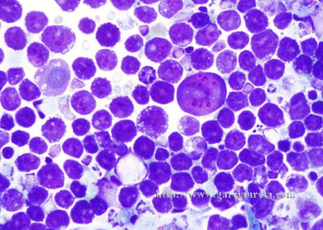
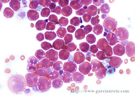
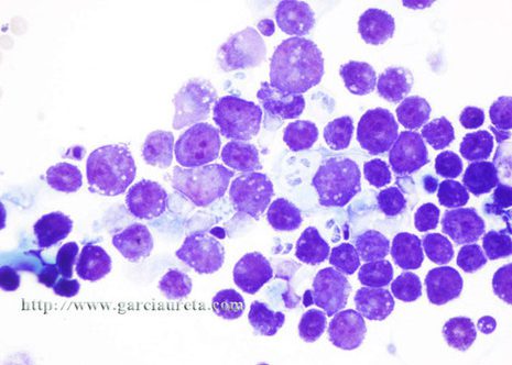
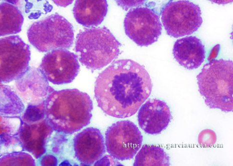
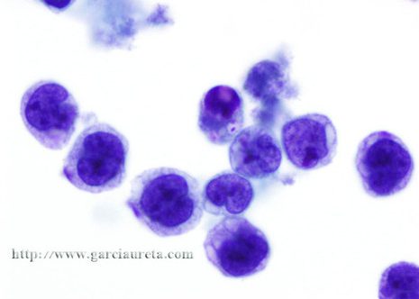
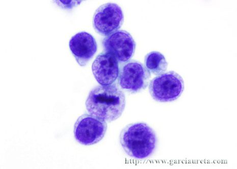
Lymphoma cells of the higher grades are generally not difficult to recognize. The cells are larger as are their nuclei which may show various degrees of polymorphism, and their nucleoli are more prominent.
Serous effusions are fairly common in patients with Hodgkin´s disease especially in the pleura cavity. The cytologic feature generally is nonspecific. Effusions due to Hodgkin´s disease are not likely contain Reed-Sternberg cells.
Myeloma cells are rarely seen in serous effusions but when they are present they are usually numerous.
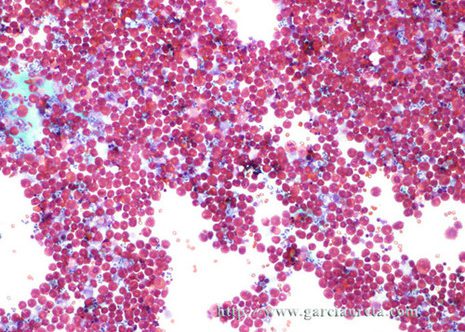
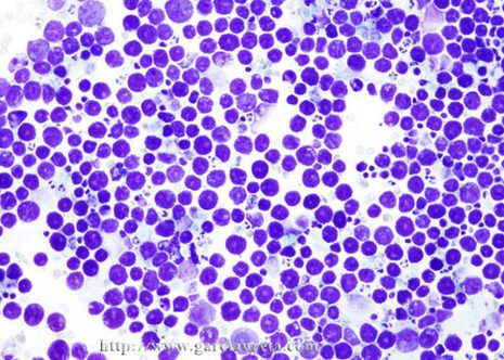
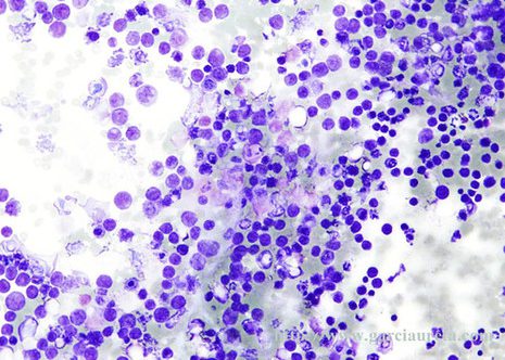
This images correspond to a 39 year old man with medical history of lymphoma and a left pleural effusion. An examination of the fluid by cytology showed large atypical lymphocytes consistent with lymphoma.
