Metastatic cancer is a far more comme cause of enlargement of peripheral lymph nodes tan malignant lymphoma particulary in patient part of age of 50.
Lymph nodes clinically suspected of metastatic malignancy constitute one of the commonest indications for FNA cytology. In patients with know, histologically proven malignancy in which enlarged nodes appear subsequently, a cytological diagnosis is usually easily obtained and further surgery merely to confirm metastasis can be avoided.
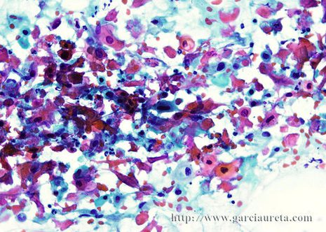
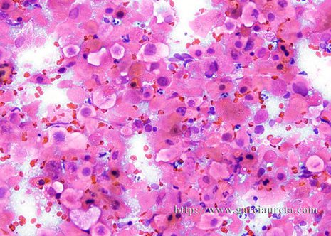
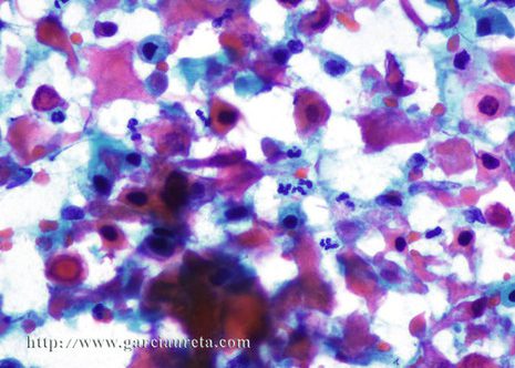
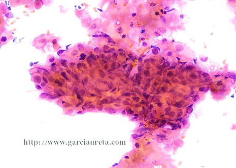
The cytologic presentation in metastatic squamous cell carcinoma depends on the degree of keratin formation by the tumor. Keratinizing cancer are readily indentified when cells with abundant sharply demarcated eosinophilic when keratinized cytoplasm and picnotic nuclei are present in smears. Keratinized squames (ghost cells) may also occur. The diagnosis of keratin-forming carcinoma is relatively easy.
Squamous cell carcinoma is the commonest type of primary carcinoma of the head and neck. The tongue is the most common site for squamous cell carcinoma within the oral cavity. Lymph node metastasis in carcinoma of the tongue is frequent.
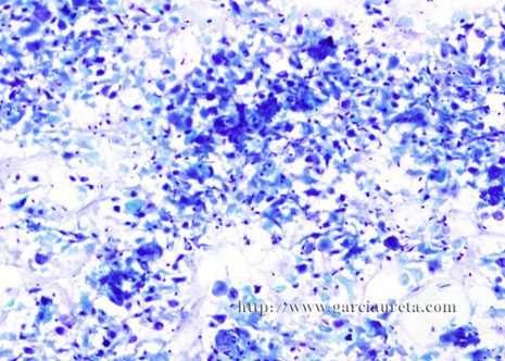
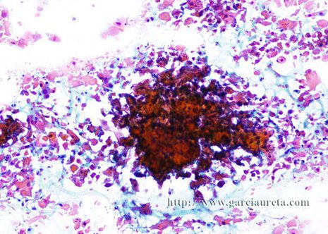
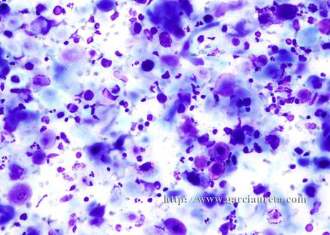
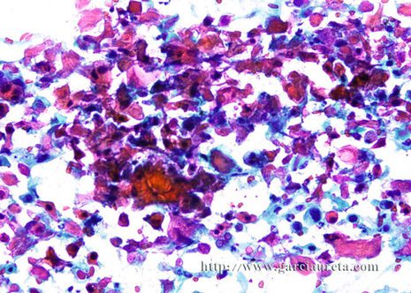
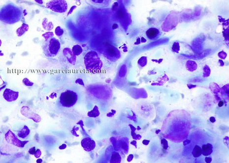
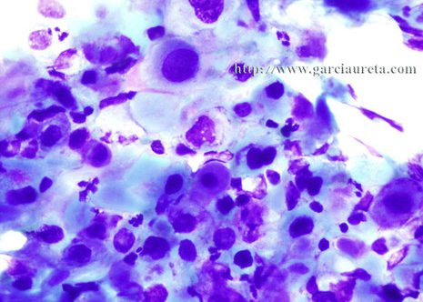
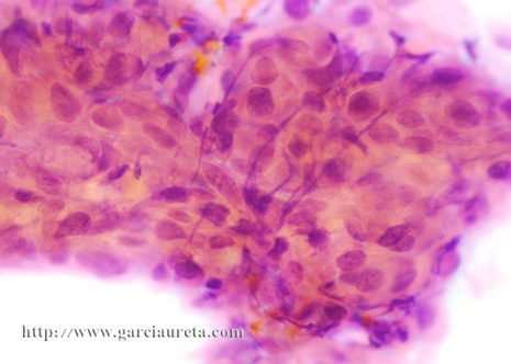
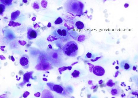
The images correspond to the FNA cytology of the cervical lymph node of a woman 81 years of age with a lesion in tongue.
The FNA smears contained abundant pleomorphic cells with dense cytoplasm and irregular nuclear shape and keratined squames. These morphologic features were interpreted as consistent with well-differentiated squamous cell carcinoma.

